
 2025/10/14
In microscopy, image quality is crucial for accurate analysis and observation. Whether you’re studying biological specimens, materials, or conducting medical research, the ability to capture detailed, high-quality images is essential. One of the key factors that determine the quality of an image in microscopy is the pixel size of the detector. Pixel size plays a significant role in light collection, which directly impacts the resolution, sensitivity, and clarity of the images produced.
What is Object Space Pixel Size in Microscopy?
Object space pixel size refers to the physical size of each pixel in the object space, or the space that the microscope is imaging. It essentially defines how much of the actual specimen each pixel in the image represents. In simple terms, smaller object space pixel sizes allow you to capture more detail from the specimen, while larger pixel sizes result in a coarser image with less detail.
The significance of object space pixel size lies in its ability to directly impact the resolution and quality of your microscopic images. High-resolution images, which are essential for accurate measurements and detailed analysis, rely on smaller object space pixel sizes. On the other hand, larger pixel sizes may compromise image quality, especially when dealing with fine structures like cells, tissues, or nanoparticles.
Figure 1: Microscope lightpath and object space pixel size definition
The object space pixel size is the width or height of the original imaging subject that is covered by a single camera pixel in the image. For microscopes, this is determined by total system magnification.
How to Calculate Object Space Pixel Size
The object space pixel size is given by:
Total magnification is given by multiplying together the magnification of all optical components in the light path.
The primary magnification in a microscope system comes from the objective lens, e.g., 10x, 20x or 60x objectives. Occasionally, there can be other magnifying lenses in the light path, including within the microscope body, or within the camera mount. It is important to check additional magnification, as lenses in camera mounts especially may not always be obvious without removing and inspecting the mount.
Measuring Magnification
In any case, it can be wise to accurately measure the total magnification of an optical system, by acquiring an image of a graticule, precise ruler or other object of known size, and looking up the camera pixel size on the camera specification sheet. Magnification of microscope objectives and other lenses can vary by a few percent from their nominal value.
Note: The 10x magnification typically added by microscope eyepieces is not included in the camera’s object space pixel size calculation.
Factors Affecting Object Space Pixel Size
Several factors influence object space pixel size in microscopy. These factors include:
● Objective Lens Magnification: The higher the magnification of the objective lens, the smaller the object space pixel size. However, increasing magnification also requires higher-quality optics to avoid blurring or distortion.
● Sensor Resolution and Pixel Size: The resolution and pixel size of the camera sensor play a critical role. A sensor with smaller pixels will yield smaller object space pixel sizes, resulting in higher resolution images.
● Optical System Setup: The optical setup, including any intermediate optics like eyepieces or beam-splitters, can influence the total magnification and, consequently, the object space pixel size.
● Camera Sensor Type (CMOS vs. CCD): The type of camera sensor used can also affect the pixel size. CMOS sensors, for instance, are commonly used in scientific applications for their efficiency and lower noise.
These factors must be carefully considered when designing your microscopy system to optimize the image quality for specific applications.
How to Measure Object Space Pixel Size and How to Change it
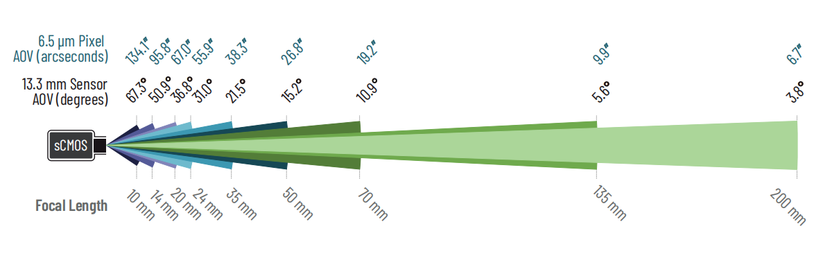
Figure 2: Angle of view at different lens focal lengths
Lens focal length determines the angle of view (AOV) of the camera sensor, and the AOV per pixel.
The specific values will depend upon the sensor size and pixel size of the camera. Example shown is for a standard 4MP sCMOS camera with a 13.3mm x 13.3mm square sensor, and 6.5 μm x 6.5 μm pixels.
For lens-based systems, the concept of object space pixel size is somewhat more complicated than for microscopes.
Microscopes have a fixed, flat focal plane that remains perpendicular to the optical axis or parallel to the camera throughout the field of view. Importantly, the optical setup of a microscope objective is usually ‘telecentric’, meaning objects that are closer to the objective do not appear larger, as if viewed without perspective. The object space pixel size is then identical throughout the field of view.
In the vast majority of lens-based systems, however, we do have to account for perspective. Combined with the larger depth of field (distance from the lens within which objects appear in focus) typical for lens-based systems, accurately defining object space pixel size can be challenging and may be different in different parts of the image.
Further, theoretical calculation of object space pixel size requires knowing both the distance from the sensor and the focal length of the lens. Given that for many lenses, the focal length can be smoothly changed between set limits (typically called ‘zoom’ lenses), the precise focal length can be challenging to ascertain.
Using angular field of view per pixel
Far simpler and more universal for lens-based systems is the angle of view per pixel, in x and y. This exhibits very similar scaling relationships to object space pixel size with regard to light collection ability and spatial sampling, but does not depend on the distance of the imaging subject to the camera. For fixed focal length lenses (also known as ‘prime’ lenses) this angular field of view per pixel is fixed for a given camera pixel size. For zoom lenses with adjustable focal length, the angle of view in x or y depends on that focal length. In both cases, the angle of view per pixel in arcseconds is closely approximated by:
Where 1 degree = 3600 arcseconds. The same formula can be used for the AOV of the sensor for long focal lengths (>50mm), with the sensor size substituted for pixel sizes. Like microscope pixel size, the light collection ability of pixel scales with the angle of view per pixel squared.
Noteworthy, however, be aware that due to geometrical constraints of lenses, the angle of view will differ subtly for pixels in different parts of the sensor, and this will depend upon the specific lens used.
Practical Applications of Adjusting Pixel Size in Microscopy
Adjusting object space pixel size in microscopy cameras has several practical applications, especially when working with intricate samples in research and diagnostics. For example:
● Live Cell Imaging: In biological microscopy, smaller pixel sizes are crucial for capturing fine details of cells, such as sub-cellular structures and organelles.
● Tissue Analysis: When examining tissue samples, adjusting pixel size allows for better resolution, enabling more accurate measurements of tissue layers and structures.
● Nanotechnology: In the study of nanoparticles and nanostructures, high-resolution imaging is essential. Smaller pixel sizes enable the detection of features that are otherwise invisible to the naked eye.
By carefully adjusting the object space pixel size, you can improve the resolution and accuracy of your measurements, leading to more reliable results.
Conclusion
Understanding how to calculate and adjust object space pixel size is essential for obtaining high-quality, detailed images in microscopy. By considering factors such as sensor resolution, objective lens magnification, and calibration techniques, you can optimize your system for precise imaging and measurements. With the right adjustments, you can ensure that your microscopy work provides the highest level of accuracy, whether you’re studying cells, tissues, or materials.
Ready to optimize your microscopy imaging system? Explore our range of microscopy accessories, cameras, and software tools to enhance your research and imaging capabilities. Contact us today to learn more about our products and how we can help improve your microscopy setup.
Tucsen Photonics Co., Ltd. All rights reserved. When citing, please acknowledge the source: www.tucsen.com


