

Application Challenges
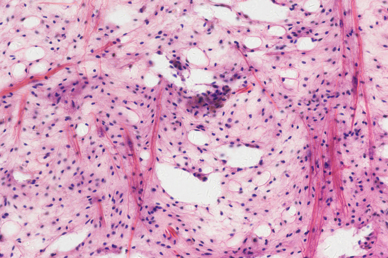
Brightfield microscopy relies on the absorption and scattering of light by biological specimens to generate contrast. It is the most classical and widely adopted imaging modality in life sciences, frequently used in pathological slide examination, histological studies, and educational demonstrations.
Biological samples span a wide range of spatial scales, from nuclei and cytoplasm to complete tissue structures, covering dimensions from micrometers to millimeters. In their unstained state, samples are nearly transparent with minimal refractive index differences, resulting in poor contrast. Consequently, various staining techniques are commonly employed to enhance image contrast. Based on these characteristics, brightfield microscopy places high demands on camera performance, particularly in spatial resolution and color fidelity.
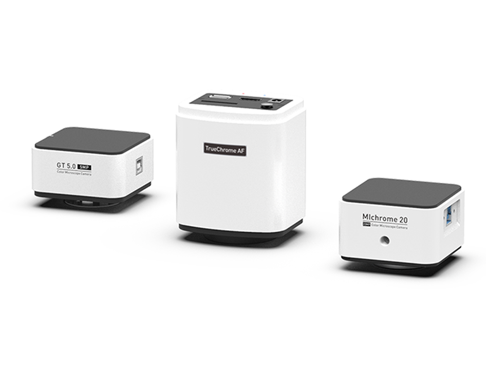
TrueChrome/ MIchrome/GT Seires
Classic Color Microscopy Cameras
Resolutions from 2 megapixels up to 20 megapixels are available. Featuring ISP (Image Signal Processing) based precise color reproduction, these cameras are paired with Tucsen’s Mosaic software specially built for microscopy users, enabling efficient imaging workflows.
TrueChrome Series (HDMI): No computer needed—captures, processing, and measurements can be done using just a mouse; higher-end options offer 4K-WiFi, autofocus, etc.
MIchrome Series (USB 3.0): Comes with Mosaic 3.0 professional edition software—allows real-time image stitching, focus stacking, and other advanced features.
GT Series (USB 2.0): Enhanced with graphics acceleration to ensure raw image output while significantly boosting frame rate; a cost-effective option.
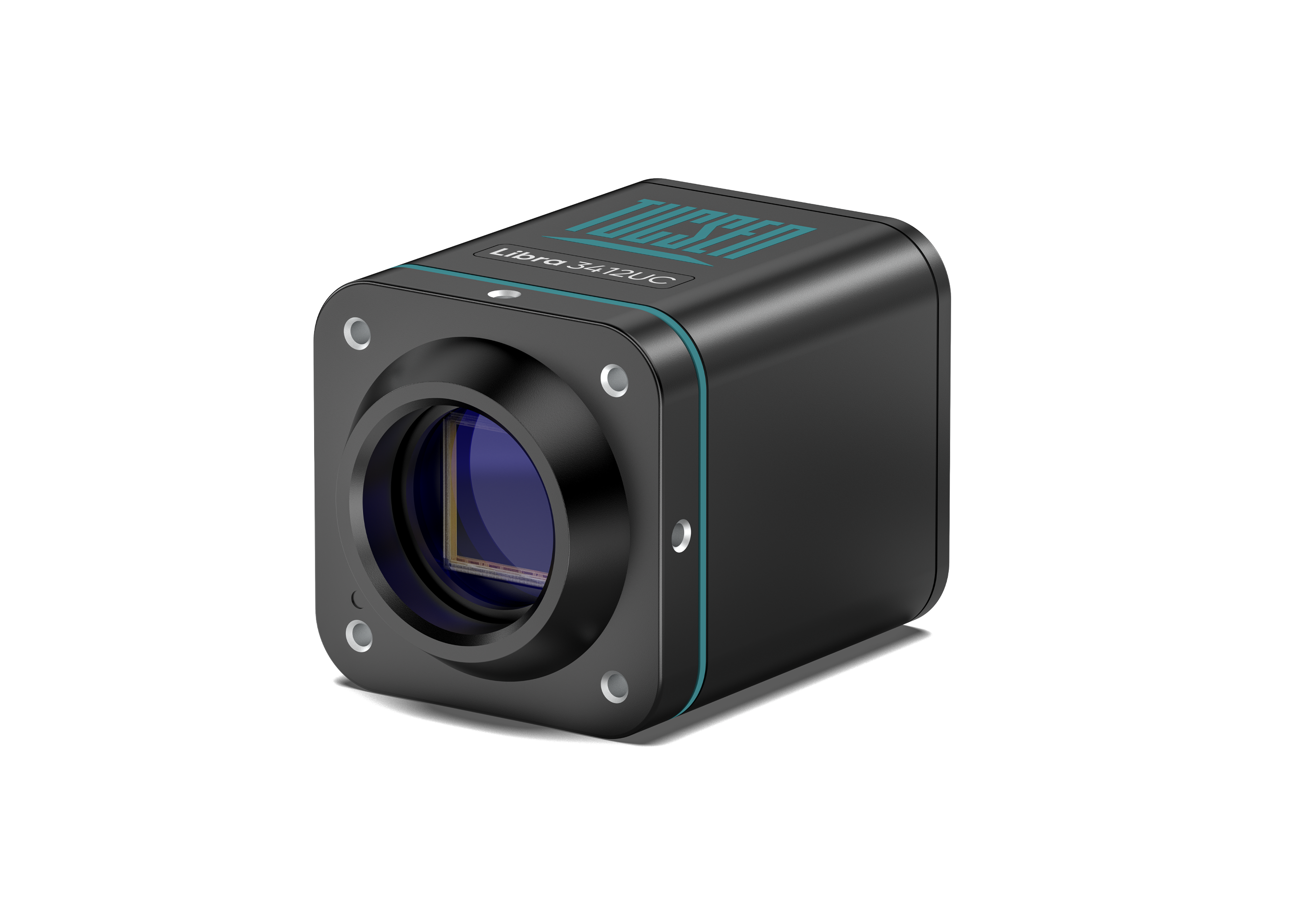
Libra 3405UC / 3412UC Coming soon
New-generation High-Performance Color Microscopy Cameras
Available in 5 MP / 12 MP resolutions, with AI color correction to provide exceptional color fidelity for pathology and related applications. Also comes with Tucsen’s Mosaic professional image processing & analysis software to enable efficient microscopy operation.
3.4 μm pixel size, better suited for optical resolution sampling under magnifications below 40×, for high detail recovery.
Broadband spectral response from 350 nm to 1100 nm, making it suitable for fluorescence and near-infrared (NIR) detection.
USB 3.0 plug-and-play interface, designed for real-time microscopy imaging workflows.


