

Application Challenges
Brightfield microscopy relies on the absorption and scattering of light by biological specimens to generate contrast. It is the most classical and widely adopted imaging modality in life sciences, frequently used in pathological slide examination, histological studies, and educational demonstrations.
Biological samples span a wide range of spatial scales, from nuclei and cytoplasm to complete tissue structures, covering dimensions from micrometers to millimeters. In their unstained state, samples are nearly transparent with minimal refractive index differences, resulting in poor contrast. Consequently, various staining techniques are commonly employed to enhance image contrast. Based on these characteristics, brightfield microscopy places high demands on camera performance, particularly in spatial resolution and color fidelity.
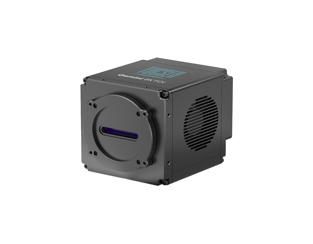
Gemini 8KTDI
High-Speed TDI-sCMOS Camera
Spectral Response: 180 nm–1100 nm, supporting UV, visible, and near-infrared applications.
TDI Stages: 256 stages, effectively enhancing SNR in low-light imaging.
Speed: 8K line frequency up to 1 MHz, doubling the overall throughput compared to the previous generation.
Cooling: Stable and reliable cooling reduces thermal noise during high-speed operation.
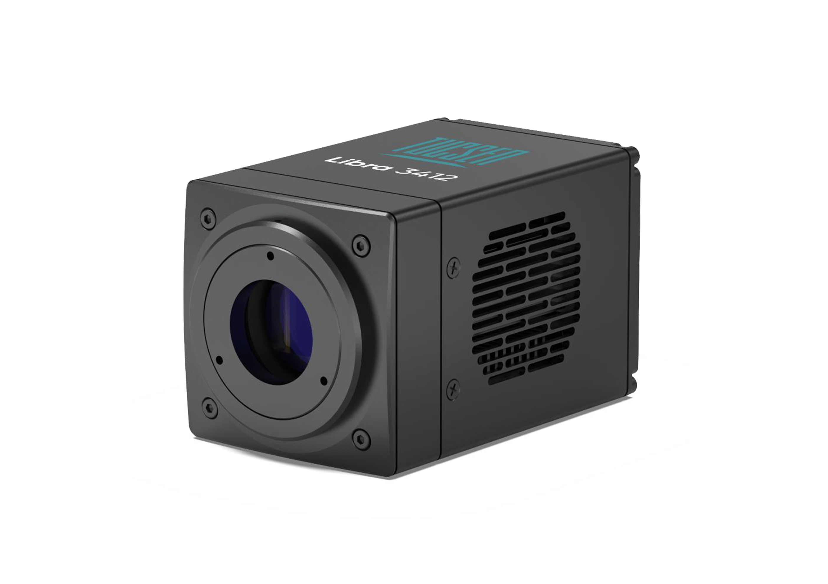
Libra 3412 Series Coming soon
Global Shutter Monochrome / Color CMOS Cameras
Spectral Response: 350 nm–1100 nm, with enhanced near-infrared response, suitable for multi-channel fluorescence imaging.
Resolution & Pixel Size: 12 MP, 3.4 µm pixels, matching optical sampling requirements below 40× magnification for high detail fidelity.
Color Features: Color models provide AI-based color correction, delivering accurate color reproduction for applications like pathology.
Interface & Speed: Color GigE high-speed interface, full-resolution frame rate up to 98 fps at 12 MP (twice the speed of USB3.0).
Cooling: Mild cooling reduces thermal interference during prolonged operation.
Compact Design: Facilitates system integration and overall performance enhancement.


