
 2025/09/30
Although color cameras dominate the consumer camera market, monochrome cameras are more common in scientific imaging.
Camera sensors are not inherently capable of detecting the color, or the wavelength, of the light they collect. Achieving a color image requires a number of compromises in sensitivity and spatial sampling. However, in many imaging applications, such as pathology, histology or some industrial inspection, color information is essential, so color scientific cameras are still commonplace.
This article explores what color scientific cameras are, how they operate, their strengths and limitations, and where they outperform their monochrome counterparts in scientific applications.
What Are Color Scientific Cameras?
A color scientific camera is a specialized imaging device that captures RGB color information with high fidelity, precision, and consistency. Unlike consumer-grade color cameras that prioritize visual appeal, scientific color cameras are engineered for quantitative imaging where color accuracy, sensor linearity, and dynamic range are crucial.
These cameras are widely used in applications such as brightfield microscopy, histology, materials analysis, and machine vision tasks where visual interpretation or color-based classification is essential. Most color scientific cameras are based on CMOS or sCMOS sensors, designed to meet the rigorous demands of scientific and industrial research.
For an in-depth look at different imaging systems, explore our selection of high-performance scientific camera models built for professional applications.
Achieving Color: The Bayer Filter
Conventionally, color detection in cameras is achieved through the same means as color reproduction on monitors and screens: through the combinations of nearby red, green and blue pixels into full-color 'superpixels'. When the R, G and B channels are all at their maximum value, a white pixel is seen.
As silicon cameras cannot detect the wavelength of incoming photons, the separation of each R, G or B wavelength channel must be achieved through filtering.
In red pixels, an individual filter is placed over the pixel to block all wavelengths but those in the red part of the spectrum, and likewise for blue and green. However, to achieve a square tiling in two dimensions despite having three color channels, a superpixel is formed from one red, one blue and two green pixels, as shown in the figure.

Bayer filter layout for color cameras
Layout of color filters added to individual pixels for color cameras using the Bayer filter layout, using repeated square 4-pixel units of Green, Red, Blue, Green pixels. Order within the 4-pixel unit can differ.
Green pixels are prioritized both because the majority of light sources (from the sun to white LEDs) exhibit their peak intensity in the green part of the spectrum, and because light detectors (from silicon-based camera sensors to our eyes) typically peak in sensitivity in the green.
When it comes to image analysis and display, however, images are not usually delivered to the user with pixels each displaying only their R, G or B value. A 3-channel RGB value is created for every pixel of the camera, through interpolating the values of nearby pixels, in a process called 'debayering'.
For example, each red pixel will generate a green value, either from the average of the four nearby green pixels, or through some other algorithm, and likewise for the four nearby blue pixels.
Pros and Cons of Color
Pros
● You can see it in color! Color conveys valuable information that enhances human interpretation, especially when analyzing biological or material samples.
● Much simpler to capture RGB color images versus taking sequential R, G, and B images using a monochrome camera
Cons
● The sensitivity of color cameras is reduced drastically compared to their monochrome counterparts, depending on wavelength. In the red and blue part of the spectrum, due to only one in four pixel filters passing these wavelengths, light collection is at most 25% that of an equivalent monochrome camera in these wavelengths. In green, the factor is 50%. In addition, no filter is perfect: the peak transmission will be less than 100%, and may be much lower depending on the exact wavelength.
● Resolution of fine details is also worsened, as sampling rates are reduced by these same factors (to 25% for R, B and to 50% for G). In the case of red pixels, with only 1 in 4 pixels capturing red light, the effective pixel size for calculating resolution is 2x larger in each dimension.
● Color cameras also invariably include an infrared (IR) filter. This is due to the ability of silicon cameras to detect some IR wavelengths invisible to the human eye, from 700nm to around 1100nm. If this IR light were not filtered out, it would affect the white balance, resulting in inaccurate color reproduction, and the image produced would not match what is seen by eye. Hence, this IR light must be filtered out, meaning that color cameras cannot be used for imaging applications, which make use of these wavelengths.
How Do Color Cameras Work?
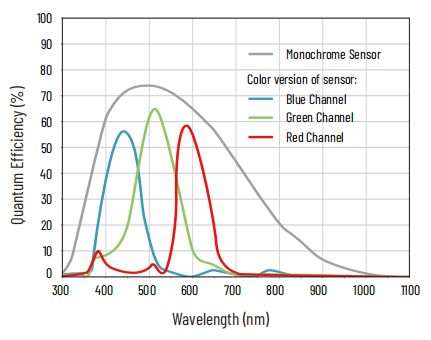
Example of a typical color camera quantum efficiency curve
Wavelength dependence of quantum efficiency shown separately for pixels with a red, blue and green filter. Also shown is the quantum efficiency of the same sensor without color filters. Addition of color filters significantly reduces quantum efficiency.
The core of a scientific color camera is its image sensor, typically a CMOS camera or sCMOS camera (scientific CMOS), equipped with a Bayer filter. The workflow from photon capture to image output involves several key steps:
1. Photon Detection: Light enters the lens and hits the sensor. Each pixel is sensitive to a specific wavelength based on the color filter it carries.
2. Charge Conversion: Photons generate an electrical charge in the photodiode beneath each pixel.
3. Readout & Amplification: Charges are converted to voltages, read out row by row, and digitized by analog-to-digital converters.
4. Color Reconstruction: The camera’s onboard processor or external software interpolates the full-color image from the filtered data using demosaicing algorithms.
5. Image Correction: Post-processing steps like flat-field correction, white balance, and noise reduction are applied to ensure accurate, reliable output.
The performance of a color camera depends heavily on its sensor technology. Modern CMOS camera sensors offer fast frame rates and low noise, while sCMOS sensors are optimized for low-light sensitivity and wide dynamic range, crucial for scientific work. These fundamentals set the stage for comparing color and monochrome cameras.
Color Cameras vs. Monochrome Cameras: Key Differences
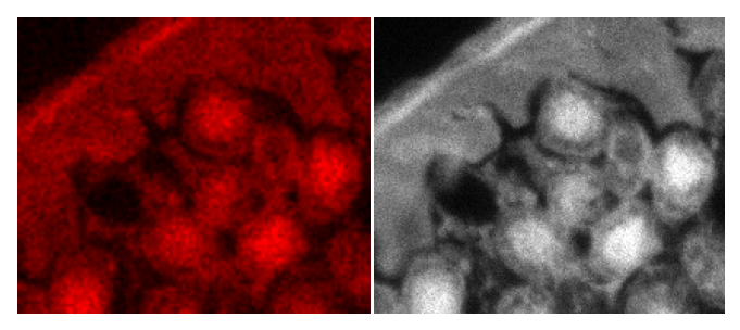
Comparison between color and monochrome camera images for low-light work
Fluorescent image with red wavelength emission detected by a color camera (left) and a monochrome camera (right), with other camera specifications remaining the same. The color image shows considerably lower signal-to-noise ratio and resolution.
While both color and monochrome cameras share many components, their differences in performance and use cases are significant. Here's a quick comparison:
|
Feature |
Color Camera |
Monochrome Camera |
|
Sensor Type |
Bayer-filtered CMOS/sCMOS |
Unfiltered CMOS/sCMOS |
|
Light Sensitivity |
Lower (due to color filters blocking light) |
Higher (no light lost to filters) |
|
Spatial Resolution |
Lower effective resolution (demosaicing) |
Full native resolution |
|
Ideal Applications |
Brightfield microscopy, histology, materials inspection |
Fluorescence, low-light imaging, high-precision measurements |
|
Color Data |
Captures full RGB information |
Captures grayscale only |
In short, color cameras are best when color matters for interpretation or analysis, while monochrome cameras are ideal for sensitivity and precision.
Where Color Cameras Excel in Scientific Applications
Despite their limitations, color cameras outperform in many specialized areas where color distinction is key. Below are a few examples of where they shine:
Life Sciences and Microscopy
Color cameras are commonly used in brightfield microscopy, especially in histological analysis. Staining techniques such as H&E or Gram staining produce color-based contrast that can only be interpreted with RGB imaging. Educational labs and pathology departments also rely on color cameras to capture realistic images of biological specimens for teaching or diagnostic use.
Materials Science and Surface Analysis
In materials research, color imaging is valuable for identifying corrosion, oxidation, coatings, and material boundaries. Color cameras help detect subtle variations in surface finish or defects that monochrome imaging might miss. For example, evaluating composite materials or printed circuit boards often requires accurate color representation.
Machine Vision and Automation
In automated inspection systems, color cameras are used for object sorting, defect detection, and labeling verification. They allow machine vision algorithms to classify parts or products based on color cues, enhancing automation accuracy in manufacturing.
Education, Documentation, and Outreach
Scientific institutions often require high-quality color images for publications, grant proposals, and outreach. A color image provides a more intuitive and visually engaging representation of scientific data, especially for interdisciplinary communication or public engagement.
Final Thoughts
Color scientific cameras serve an essential role in modern imaging workflows where color differentiation is important. While they may not match monochrome cameras in sensitivity or raw resolution, their ability to deliver natural, interpretable images makes them indispensable in fields ranging from life sciences to industrial inspection.
When selecting between color and monochrome, consider your imaging goals. If your application requires low-light performance, high sensitivity, or fluorescence detection, a monochrome scientific camera might be your best option. But for brightfield imaging, material analysis, or any task involving color-coded information, a color solution may be ideal.
To explore advanced color imaging systems for scientific research, browse our full lineup of high-performance CMOS cameras and sCMOS models tailored to your needs.
Tucsen Photonics Co., Ltd. All rights reserved. When citing, please acknowledge the source: www.tucsen.com


