Abstract
In order to study the localization and orientation of proteins in subcellular structures, peng Xi and his colleagues developed polarized structured light microscopy (pSIM). The study is in the journal Nature Communications.
This work "empowers" the existing SIM system by deeply exploring the potential characteristics of SIM technology and commercial instruments, and excavates the inherent polarization detection characteristics of the existing SIM system that even its inventors have not noticed, so that the existing system can realize the function of polarization SIM without any modification. This enables many life science laboratories with SIM systems to directly analyze polarization SIM, which will greatly advance the research of polarization super-resolution imaging.

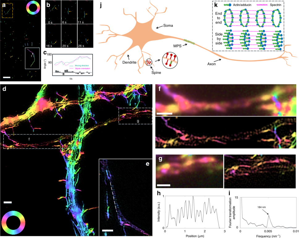
Fig.1Imaging the orientation of short actin filaments. a Dynamic imaging of the myosin-driven movement of phalloidin-labeled actin filament. The white box contains the trajectory of a short actin filament. b Magnified view of the yellow boxed region in a. The dipole orientations of the actin filaments change as they move. c Time-lapse orientation and position of the fragmented actin in b. d 2D-pSIM imaging of the actin filaments in hippocampal neurons, which clearly distinguishes between the continuous long actin filaments in the dendrite and the region of discrete actin ring structure in the axon. e Another illustration of the actin ring structure in the axon. f, g Magnified views of the boxed regions in d, which compare the results of PM and pSIM imaging. h The intensity profile of the line indicated in e, whose Fourier transform (i) shows a 184 nm periodicity, consistent with previously reported results. j, k The actin ring structure is critical to the membrane-associated periodic skeleton (MPS) in neurons. The previous model assumes an end-to-end organization for the adducin-capped actin filaments. However, pSIM reveals that the orientation of the short actin filaments is parallel to the axon shaft, supporting a side-by-side organization for the actin ring structures. Scale bars: a 2 μm and d–g 1 μm
Analysis of imaging technology
QSIM super-resolution imaging optical system is a typical low-light imaging system. The TUCSEN Dhyana 400BSI camera has a peak quantum efficiency of 95% and readout noise as low as 1.2e-, which can help the low-light system to obtain high signal-to-noise ratio imaging images. The pixel of 6.5 μm is suitable for 60x high-power objective lens. It can give full play to the advantages of lens resolution and help the system to obtain high-resolution images with clear details.
Reference source
Zhanghao K, Chen X, Liu W, Li M, Liu Y, Wang Y, Luo S, Wang X, Shan C, Xie H, Gao J, Chen X, Jin D, Li X, Zhang Y, Dai Q, Xi P. Super-resolution imaging of fluorescent dipoles via polarized structured illumination microscopy. Nat Commun. 2019 Oct 16;10(1):4694. doi: 10.1038/s41467-019-12681-w. PMID: 31619676; PMCID: PMC6795901.

 22/03/03


