

Application Challenges
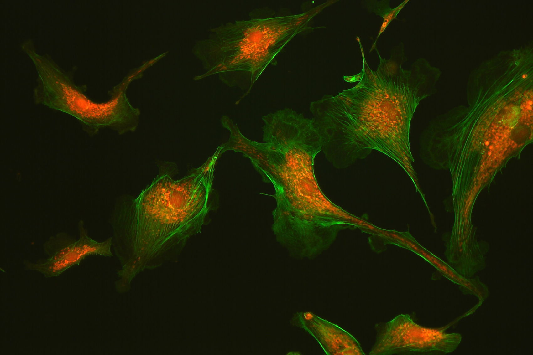
Confocal microscopy employs point scanning and a pinhole aperture to effectively suppress out-of-focus light, producing high-contrast optical section images that can be reconstructed into three-dimensional datasets. It is a powerful tool for imaging thick tissue slices and 3D cellular structures, allowing researchers to explore spatial relationships and cellular distributions.
However, traditional point-scanning systems rely on photomultiplier tubes for fluorescence detection, which limits acquisition speed and the ability to capture fast dynamic events. To address these limitations, spinning-disk confocal microscopy was developed. By employing multiple rapidly scanning laser beams and a rotating pinhole disk, spinning-disk systems achieve high-speed optical sectioning while detecting fluorescence with cameras such as sCMOS or EMCCD. This approach enables simultaneous acquisition of large fields of view with minimal phototoxicity, making it particularly suitable for live-cell imaging and rapid 3D experiments.
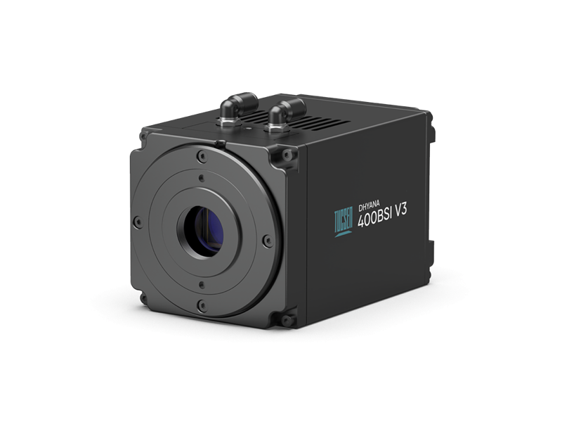
Dhyana 400BSI V3
Classic 6.5 µm BSI sCMOS Camera
Pixel Size: 6.5 µm, optimized for 40×–60× high-NA objectives.
Shutter Modes: Multiple rolling shutter modes, tailored for scanning and light-sheet imaging.
Calibration: PRNU/DSNU correction ensures uniform background for accurate quantitative analysis.
Interface: USB 3.0 and Camera Link.
Cooling: Water + air cooling design for stable, low-noise operation.
Compact Design: Lightweight at 995 g, low power consumption of 45 W.
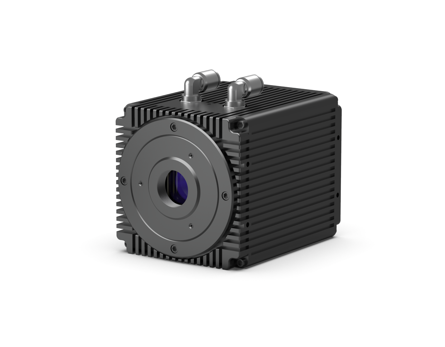
Dhyana 95 V2
Classic 11 µm BSI sCMOS Camera
Pixel Size: 11 µm, ideal for Nyquist sampling with 60×–100× high-NA objectives.
Sensor Area: Large 32 mm imaging area for extended field-of-view imaging.
Full-Well Capacity: High, supporting quantitative measurements with a wide dynamic range.
Interface: Dual options – USB 3.0 and Camera Link.
Cooling: Hybrid water + air cooling system effectively suppresses dark current.
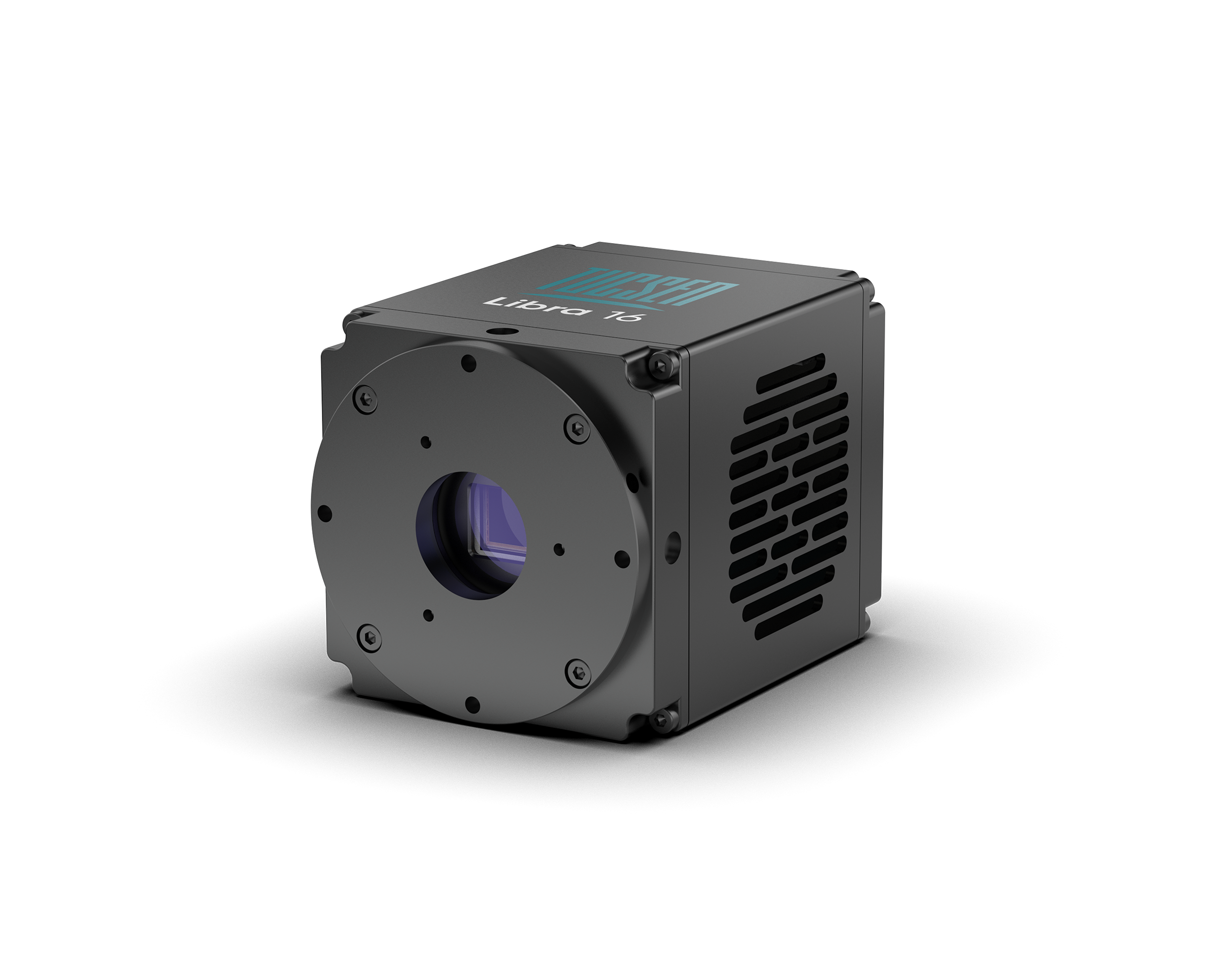
Aries 16
16 μm Large-Pixel BSI sCMOS Camera
16 μm large pixels provide ~6× higher photon collection efficiency than 6.5 μm pixels, greatly enhancing weak-light sensitivityUltra-low readout noise (~0.9 e⁻) and up to 90% quantum efficiency, enabling single-photon detection
Deep cooling up to 60°C below ambient effectively reduces dark current and improves SNR
High full-well capacity (~74 ke⁻) allows simultaneous measurement of strong and weak signals in complex light fields
HDR & low-noise readout modes support flexible switching between high-dynamic and weak-light imaging scenarios
Reliable and stable cooling minimizes data drift and improves measurement accuracy


