

Application Challenges
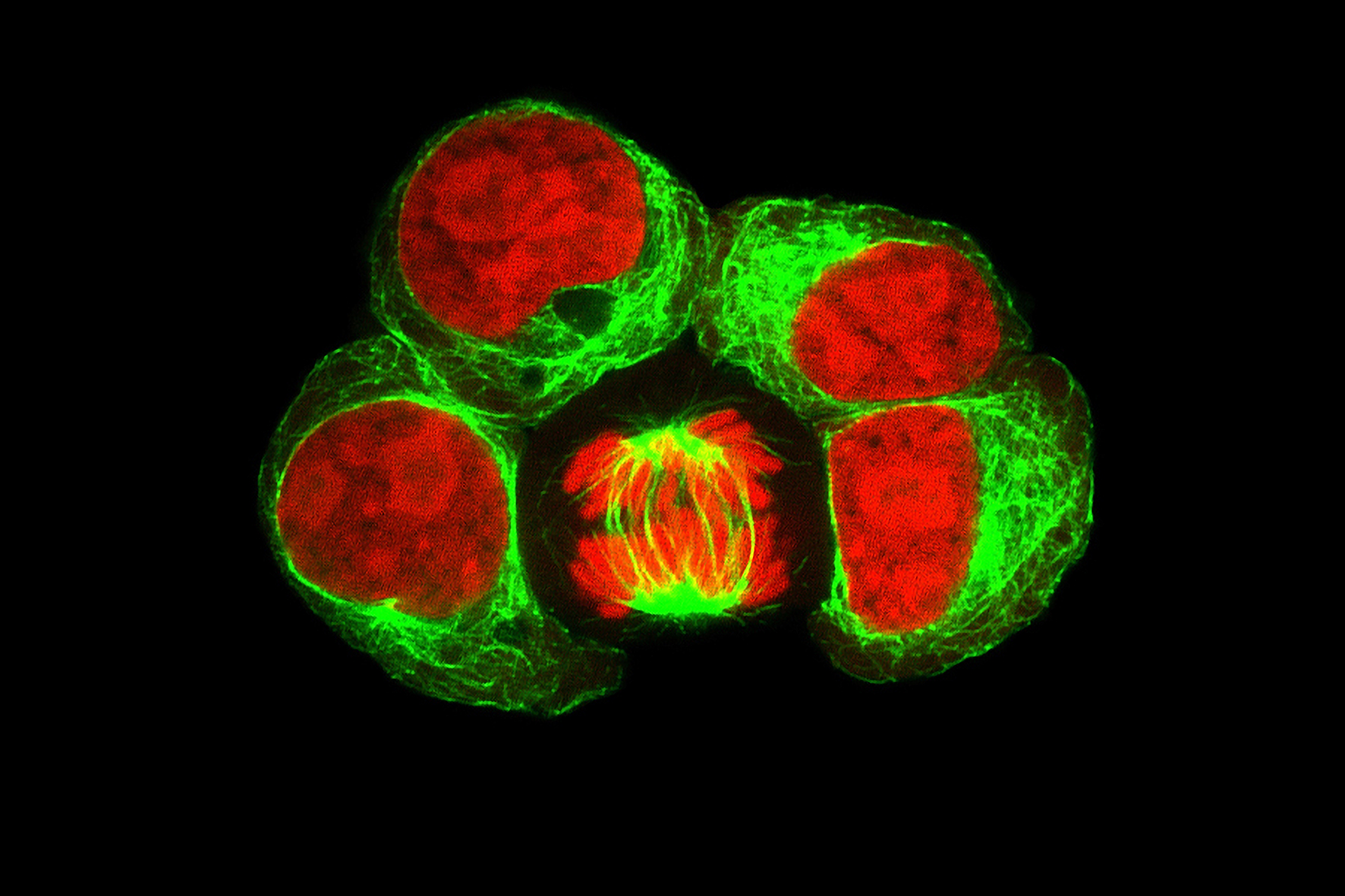
Live-cell imaging captures cellular behaviors and functions under physiologically relevant conditions, including migration, division, signaling pathway activation, and subcellular structural changes. It provides critical tools for understanding cellular functions, disease mechanisms, and drug effects, and is widely applied in both basic life sciences research and pharmaceutical development.
The primary challenges include the dynamic nature of live cells, weak signal intensities, and environmental sensitivity to temperature, CO₂ concentration, and humidity. These challenges require cameras with high sensitivity to detect faint signals, low noise to ensure image quality, and high frame rates to record rapid dynamic events, ensuring reliable and reproducible live-cell observations.
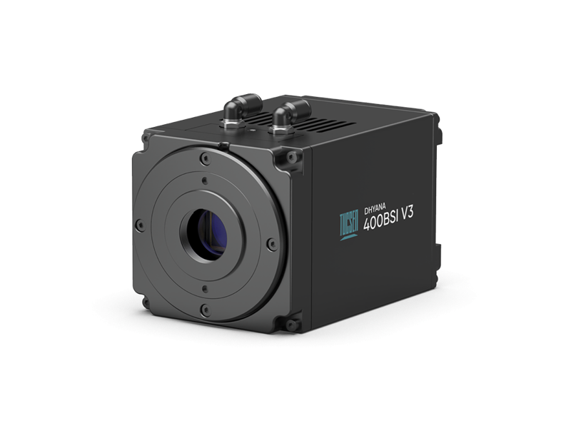
Dhyana 400BSI V3
Classic 6.5 µm BSI sCMOS Camera
Pixel Size: 6.5 µm, optimized for 40×–60× high-NA objectives.
Shutter Modes: Multiple rolling shutter modes, tailored for scanning and light-sheet imaging.
Calibration: PRNU/DSNU correction ensures uniform background for accurate quantitative analysis.
Interface: USB 3.0 and Camera Link.
Cooling: Water + air cooling design for stable, low-noise operation.
Compact Design: Lightweight at 995 g, low power consumption of 45 W.
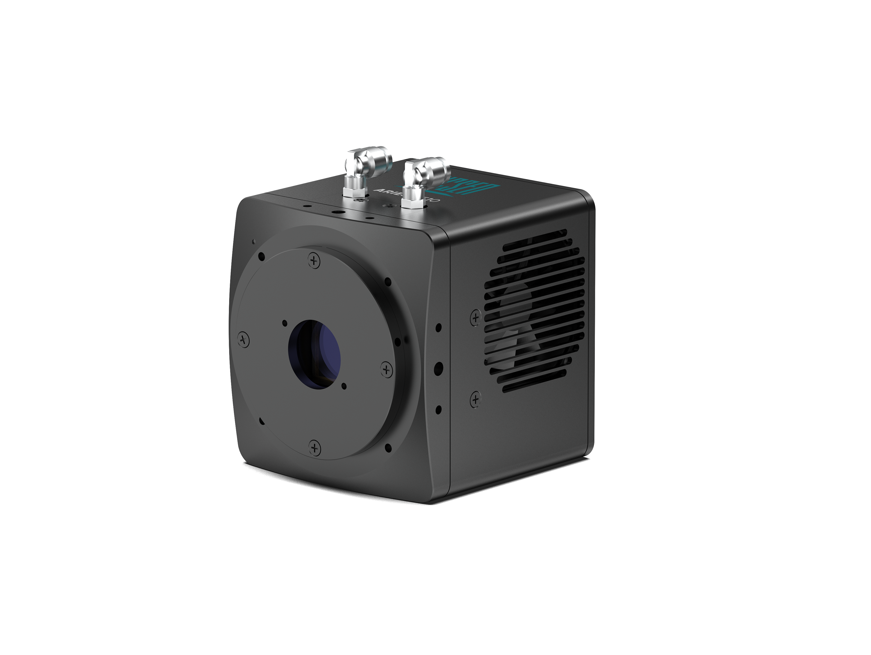
Aries 6510
Large-Format 6.5 µm BSI sCMOS Camera
Quantum Efficiency: Peak QE up to 95%, readout noise <0.7 e⁻, capable of single-photon measurements.
Pixel Size: 6.5 µm, suitable for Nyquist sampling with 40×–60× high-NA objectives.
Sensor Size: 29.4 mm, 10.2 MP full-resolution frame rate up to 150 fps, enabling high-throughput performance.
Interface: High-speed GigE interface, lossless data transmission, flexible cabling.
Cooling: Highly reliable cooling reduces data fluctuation and improves measurement precision.
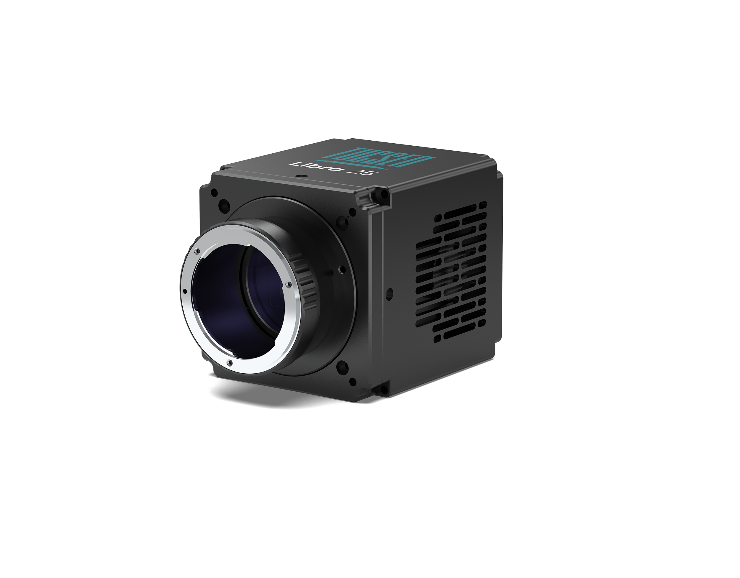
Libra 25
Large FOV Monochrome Cooled CMOS Camera
Large FOV: 25 mm sensor, compatible with wide-field microscope optical systems.
High Quantum Efficiency: Up to 92% QE, readout noise as low as 1.0 e⁻, supporting high SNR imaging applications.
Pixel Sizes:7.5 µm pixels, matched to 60× objectives for low-light imaging.
Trigger Interface: Supports synchronization with illumination devices for high-speed multi-channel imaging experiments.
Software Compatibility: Compatible with Mosaic 3.0 software, enabling real-time quantitative analysis and intelligent image processing.


