

Application Challenges
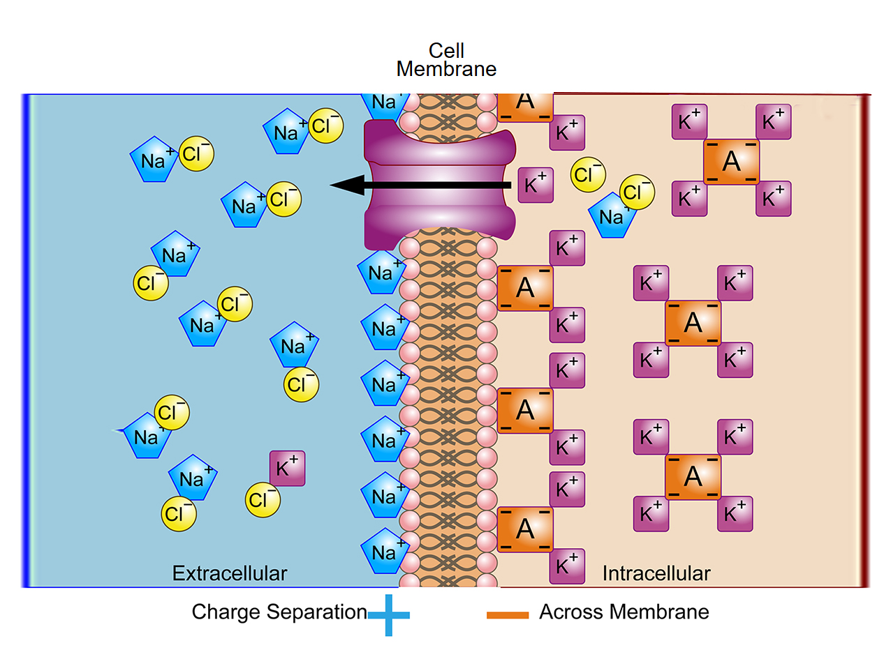
Membrane potential imaging uses fluorescent voltage-sensitive dyes or genetically encoded voltage indicators (GEVIs) to monitor dynamic changes in cell membrane potential. Depolarization or repolarization induces rapid changes in probe fluorescence intensity or spectrum, enabling optical detection of millisecond-scale electrical signals. This technique provides a noninvasive method for observing electrical activity of single cells and neural networks.
such experiments are typically performed with widefield fluorescence, two-photon, light-sheet, or other high-speed imaging platforms. They require subcellular spatial precision while detecting fluorescence changes of only a few percent (ΔF/F), corresponding to rapid membrane depolarization and repolarization events on the millisecond timescale. Consequently, imaging systems must combine ultra-high temporal resolution excellent signal-to-noise performance, and photostability to minimize phototoxicity during prolonged recordings of excitable cells and neural networks.
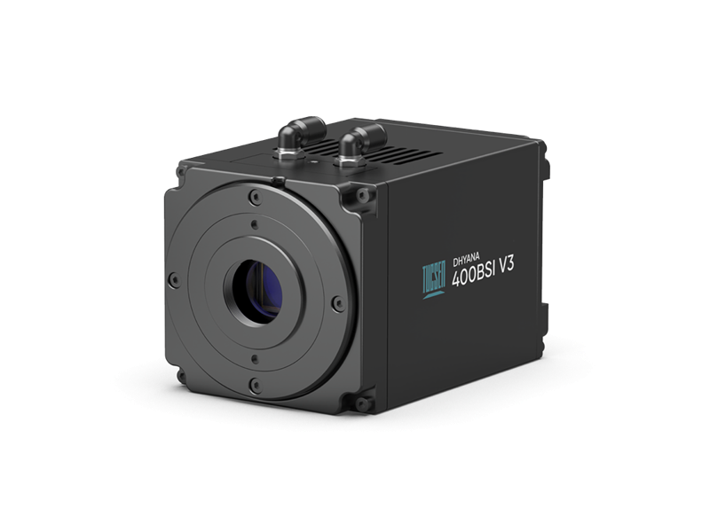
Dhyana 400BSI V3
Classic 6.5 µm BSI sCMOS Camera
Pixel Size: 6.5 µm, optimized for 40×–60× high-NA objectives.
Shutter Modes: Multiple rolling shutter modes, tailored for scanning and light-sheet imaging.
Calibration: PRNU/DSNU correction ensures uniform background for accurate quantitative analysis.
Interface: USB 3.0 and Camera Link.
Cooling: Water + air cooling design for stable, low-noise operation.
Compact Design: Lightweight at 995 g, low power consumption of 45 W.
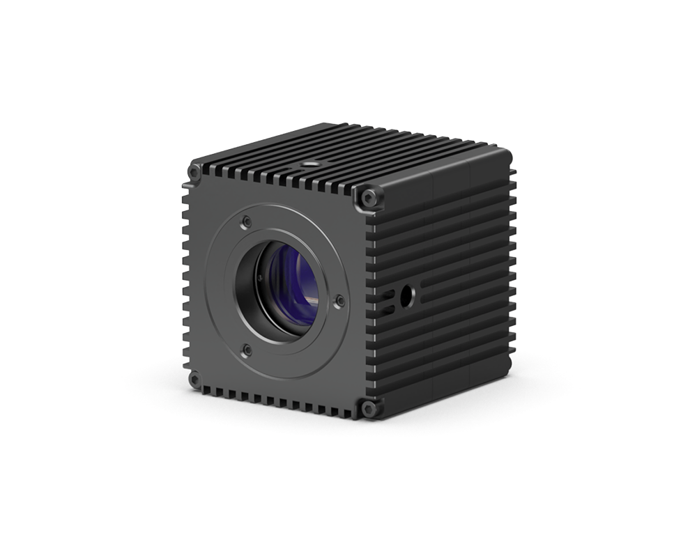
Dhyana 401D
Compact 6.5 µm sCMOS Camera
Compact design ideal for system integration
No fan cooling, relying on excellent structural and circuit design to reduce noise, making it very suitable for live-cell experiments
External trigger function for remote experiments, reducing human interference
6.5 µm pixel size balances resolution and sensitivity, fully utilizing the optical system's resolving power
Imaging field covers the full field of view of mainstream microscopes


