
 2025/10/29
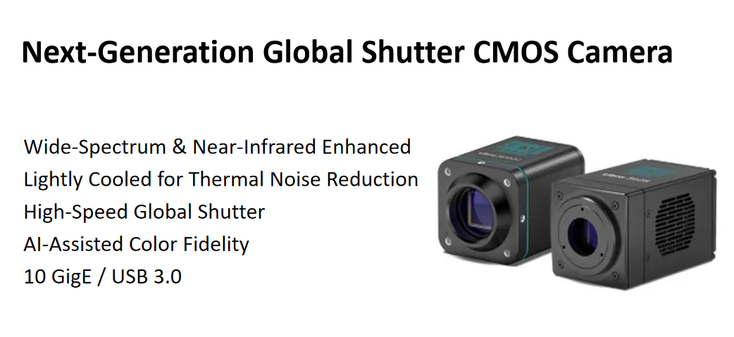
Fuzhou, China — September 2025 — Tucsen Photonics today announced the launch of the Libra 3405/3412 series, a new generation of global shutter CMOS cameras engineered to accelerate digital pathology and whole slide imaging (WSI) system upgrades. Combining high-throughput performance, AI-driven color accuracy, and cost-effective integration, the Libra 3405/3412 series sets a new standard for high-speed, high-fidelity imaging in medical diagnostics and life science research.
Empowering Next-Generation Digital Pathology
Over the past two decades, whole slide imaging (WSI)—also known as digital pathology—has evolved from early brightfield microscopy digitization into today’s AI-assisted multimodal imaging platforms. As digital pathology transitions toward higher throughput and intelligence, imaging systems face mounting challenges in balancing speed, resolution, and cost-efficiency.
The Tucsen Libra 3405/3412 series directly addresses these challenges, offering an optimized blend of imaging performance and affordability to support both system integrators and medical device manufacturers in achieving the next stage of intelligent pathology imaging.
Key Innovations of the Libra 3405/3412 Series
1. Broad Spectrum Sensitivity: Expanding Multichannel Imaging
The Libra 3405/3412 cameras deliver a broad spectral response from 350 to 1100 nm, enabling both standard DAPI–FITC–Cy3–Cy5 fluorescence imaging and advanced near-infrared (NIR) applications.
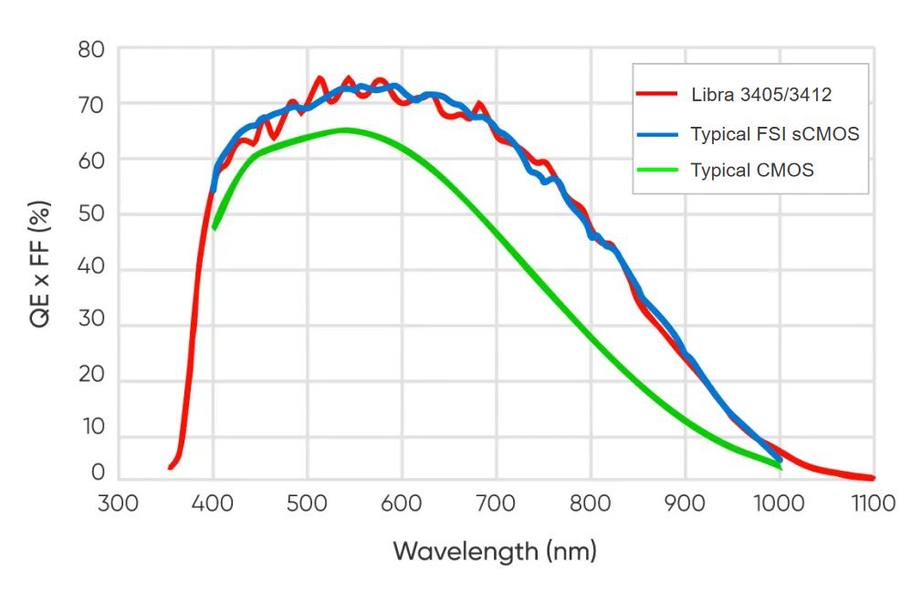
Figure 1-1. QE Curve Fitting Chart
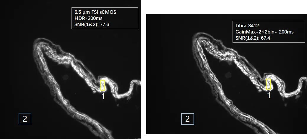
Figure 1-2. Image SNR Comparison: Libra 3412M vs. FSI sCMOS
At the critical 800 nm NIR wavelength, the Libra 3405/3412 monochrome models reach a quantum efficiency of up to 47%—approximately twice that of conventional CMOS cameras used in digital pathology—creating greater potential for six- to eight-channel imaging applications.
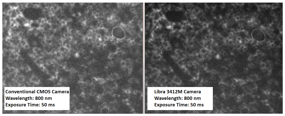
Figure 1-3. Near-Infrared Imaging Comparison: Libra 3412M vs. Conventional CMOS
Additionally, the GigE version of the Libra 3405/3412 series incorporates mild sensor cooling. This ensures exceptionally high SNR even during long-exposure imaging, providing comprehensive performance assurance for pathology system integration and extended imaging workflows.
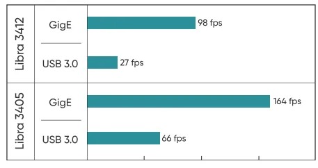
2. 10 GigE + Global Shutter: High-Speed, Artifact-Free Imaging
Equipped with a 10 GigE high-speed interface, the Libra 3405/3412 achieves frame rates up to 98 fps (12 MP) and 164 fps (5 MP)—three times faster than USB 3.0 models.
The global shutter enables true “fly-by” scanning for automated WSI, ensuring precise color alignment and eliminating motion artifacts.
Native support for Ethernet protocols further enhances remote access, data sharing, and network-based automation, paving the way for connected, high-throughput pathology systems.
3. 3.4 µm Pixels: Matched to Pathology Optics
Designed around typical pathology objectives (10×–40×), the Libra 3405/3412 employs 3.4 µm high-resolution pixels, perfectly aligned with Nyquist sampling for submicron tissue structures.
Figure 3-1. Ideal Pixel Reference for 4–100X Optical System
According to the Nyquist sampling theorem, the ideal pixel size of a camera should be approximately one-half to one-third of the product of optical resolution and magnification. As illustrated in Figure 3-2, the Libra 3405/3412 series utilizes 3.4 μm pixels, which are optimally suited for sampling in optical systems below 40× magnification. Compared with larger pixels such as 6.5 μm, this finer pixel pitch enables more detailed imaging of cellular and tissue morphology, ensuring higher precision and clarity in digital pathology workflows.

Figure 3-2. Image Resolution Comparison (6.5 μm vs. 3.4 μm)
4. AI-Powered Color Correction: Exceptional True-to-Life Color
Brightfield pathology slides typically rely on staining techniques such as H&E for diagnosis and analysis, requiring extremely high color fidelity, particularly for closely related hues such as blue, purple, and pink.
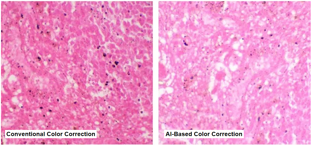
Figure 4. Conventional Color Correction vs. AI-Based Color Correction
Left: produces overly saturated colors
Right: achieves colors closer to the true view through the eyepiece
The Libra 3405/3412 series incorporates a proprietary AI-based color restoration algorithm trained specifically for brightfield pathology imaging. It eliminates the need for manual white balance adjustments by automatically adapting to color temperature and sample characteristics, ensuring accurate, true-to-eye color reproduction consistent with what pathologists observe through the microscope.
5. Optimized for Cost and Integration
Balancing sCMOS-grade imaging performance with significant cost advantages, the Libra 3405/3412 series is available in 5 MP and 12 MP, color and monochrome models, and 10 GigE or USB 3.0 interface options.
Each model is supported by Tucsen’s comprehensive SDK, ensuring rapid OEM integration and compatibility with established pathology and microscopy software ecosystems.
With nearly 20 years of OEM/ODM experience, Tucsen provides full lifecycle technical support—covering system integration, performance validation, and long-term maintenance—to help partners accelerate development and ensure product reliability.
Availability
The Tucsen Libra 3405/3412 series is now available worldwide. For technical specifications, evaluation samples, or integration support, please visit www.tucsen.com or contact Tucsen’s application engineering team.
About Tucsen Photonics
Tucsen Photonics Co., Ltd. is a leading developer and manufacturer of high-performance imaging solutions for scientific, industrial, and life science applications. With operations in China, Singapore, the United Kingdom, the United States, and Europe, Tucsen delivers innovative and reliable imaging technologies that empower researchers and engineers worldwide to see beyond the limits of vision.
Media Contact
Yuki Tang
Marketing Director
Email: yukitan@tucsen.com
Tucsen Photonics
LinkedIn: www.linkedin.com/company/tucsen
Web: www.tucsen.com
Tucsen Photonics Co., Ltd. All rights reserved. When citing, please acknowledge the source: www.tucsen.com


