
 2025/12/18
“The Aries 6510 provides the high speed, sensitivity, and resolution needed for our voltage imaging experiments. Its performance allows us to capture millisecond-scale neuronal dynamics across large fields of view for real-time imaging and analysis.”
- Eric Lowet, Erasmus MC
Group Research Aims
The Lowet lab's work develops and applies cutting-edge optical voltage imaging to study how neurons behave in awake, behaving animals. He uses genetically encoded voltage indicators (GEVIs) together with high-speed microscopy (kHz), targeted illumination, and confocal gating to improve signal-to-noise, reduce background, and image large populations and deep tissues. The lab explores how neurons switch between firing single spikes versus bursts, how those modes relate to brain rhythms during spatial navigation, and how deep brain stimulation impacts membrane voltage and network encoding. His methods allow resolving both action potentials and sub-threshold voltage dynamics optically.
The group’s goal is to link microscopic neural activity with macroscopic brain function, revealing how distributed networks change across states and between sexes. Through continuous innovation in imaging and data analysis, the lab strives to push the boundaries of systems neuroscience and brain-wide mapping.
Equipment & Experiment
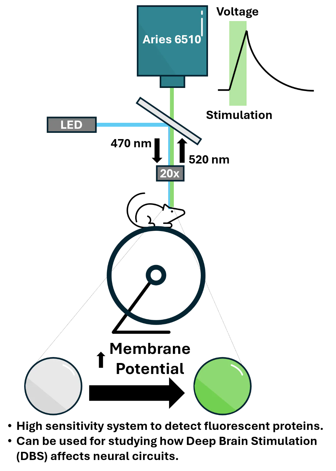
The experiment captures rapid membrane voltage in awake mouse hippocampal interneurons using a genetically-encoded voltage indicator (GEVI) using widefield fluorescence imaging with targeted illumination which is patterned via a digital micromirror device (DMD). Excitation patterns are restricted to selected cells to enhance contrast & minimise background signals. Fluorescence signals corresponding to sub-millisecond membrane potential fluctuations are recorded continuously at kilohertz frame rates.
The Tucsen Aries 6510 complements this process by providing the high speed, low noise, and sensitivity needed to resolve single action potentials across large neuronal populations. Using the camera's high sensitivity mode & binning to 13 μm or more, the group can capture subtle action potential dynamics from stimulated neurons & seizure data.
Experience with Tucsen
The experiment captures rapid membrane voltage in awake mouse hippocampal interneurons using a genetically-encoded voltage indicator (GEVI) using widefield fluorescence imaging with targeted illumination which is patterned via a digital micromirror device (DMD). Excitation patterns are restricted to selected cells to enhance contrast & minimise background signals. Fluorescence signals corresponding to sub-millisecond membrane potential fluctuations are recorded continuously at kilohertz frame rates.
The Tucsen Aries 6510 complements this process by providing the high speed, low noise, and sensitivity needed to resolve single action potentials across large neuronal populations. Using the camera's high sensitivity mode & binning to 13 μm or more, the group can capture subtle action potential dynamics from stimulated neurons & seizure data.
– Eric Lowet, Erasmus MC
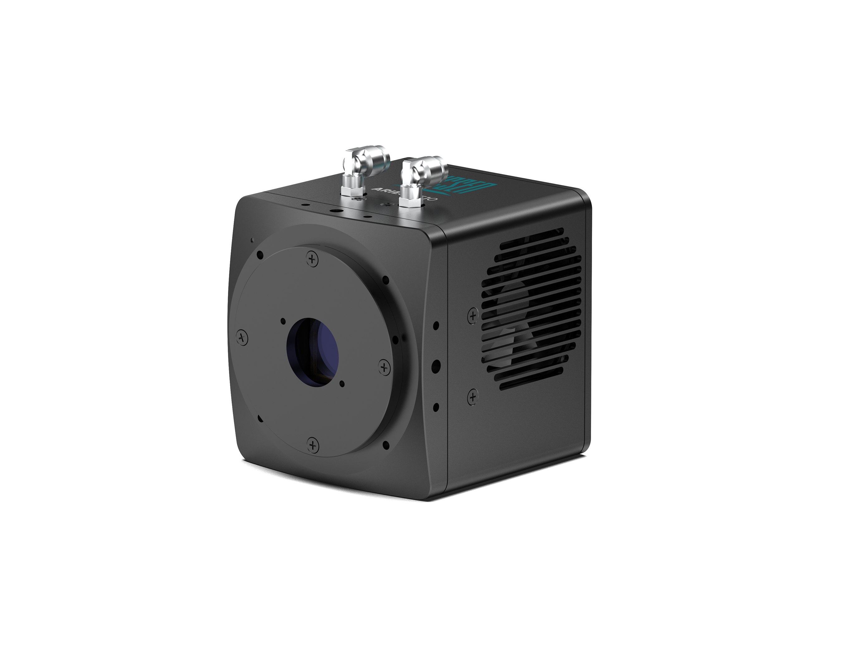
Aries 6510
The Aries 6510 achieves perfect combination of sensitivity, large FOV and high-speed performance.
● 95% Peak QE
● 150 fps @ Full Resolution
● 0.7 e- Read Noise
● 10.2 Million Pixels
● 6.5 Micron Pixels
● USB 3.0 and GigE


