
 2025/11/28
Overview
Modern neuroscience relies on the ability to capture neuronal and network activity at millisecond timescales with sufficient temporal resolution, spatial resolution, and signal-to-noise ratio. Whether in calcium imaging, voltage imaging, optogenetically coupled imaging, multiphoton deep-tissue imaging, or freely moving in-vivo preparations, researchers face the same challenges: neural signals are both fast and low in amplitude, and the required imaging window is often wide and complex. In these experimental configurations, the performance ceiling is frequently set by the detector at the end of the signal chain.
Over the past decade, sCMOS technology has demonstrated strong capability in handling weak and complex neural signals due to its high sensitivity and large field of view. At the same time, it has revealed new performance bottlenecks and further expanded the demand for next-generation detectors.
The need for higher-performance imaging detectors in neuroscience continues to grow.
Application Advantages of the Aries 6504 sCMOS Camera for Neuroscience Imaging
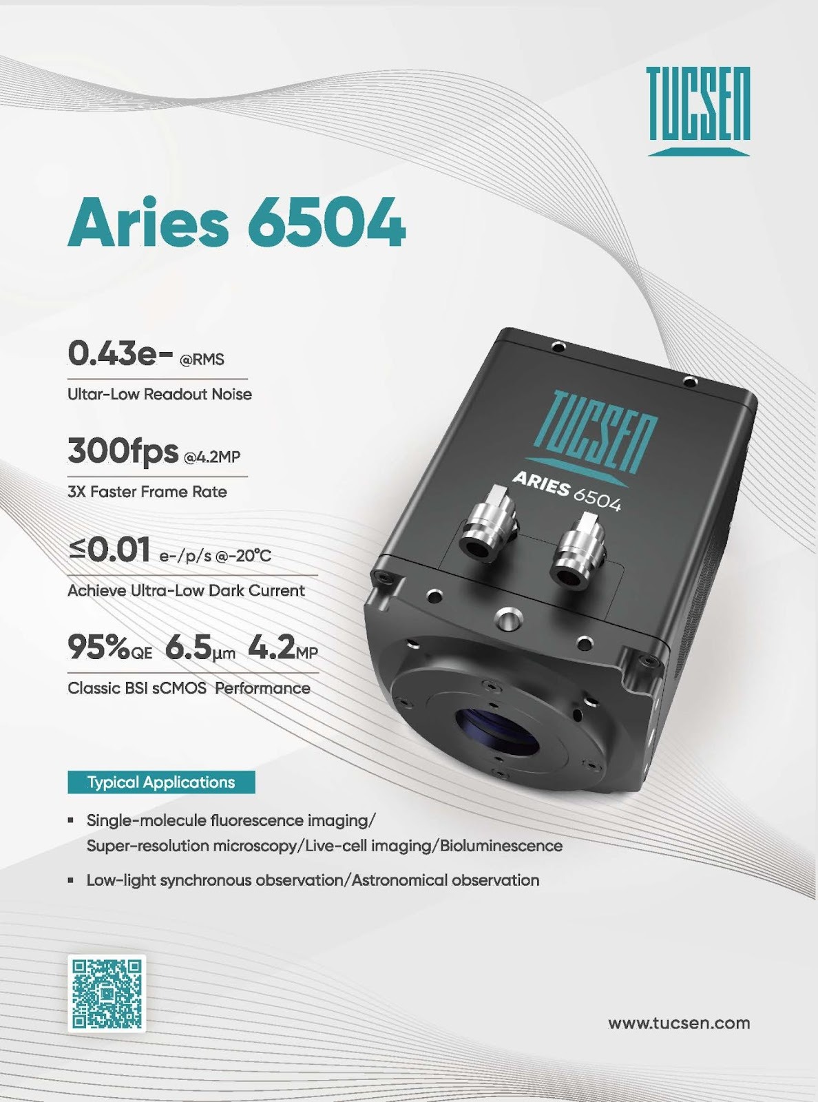
The Aries 6504 is Tucsen’s next-generation backside-illuminated sCMOS camera. Building on the classic performance of the previous-generation 6.5-μm-pixel sCMOS platform—featuring 95% peak quantum efficiency, 4-megapixel resolution, and high dynamic range—the camera delivers substantial improvements in three core performance metrics: read noise, frame rate, and dark current. These advancements enable higher-precision acquisition for high-speed, dynamic neuroscience imaging.
300 fps @ 4.2 MP Full Resolution — 3× Frame-Rate
Enabling High-Speed Voltage and Calcium Imaging Across Large Fields of View
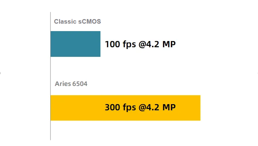
Although modern sCMOS sensors overcome the inherent speed–noise trade-offs of CCD/EMCCD technologies, recording ultrafast and transient neural activity—such as epileptiform bursts, high-frequency oscillations, or synchronous firing—still often requires ROI cropping, forcing researchers to sacrifice field of view to achieve higher frame rates. This remains a challenge for sampling rates in the hundreds to >1,000 Hz range. In addition, genetically encoded voltage indicators typically exhibit <10% ΔF/F and millisecond kinetics, demanding simultaneously high speed and low noise.
The Aries 6504 achieves 300 fps at 4.2-megapixel full resolution, representing a 3× increase over previous-generation BSI sCMOS cameras. This significantly expands the operational envelope of “high-frame-rate × large-FOV” imaging. The improvement enhances the ability to capture fast network-scale activity and supports the transition of large-scale voltage imaging from exploratory research to routine application. High frame rates also reduce temporal uncertainty in fast calcium indicators (e.g., jGCaMP8f), improving spike inference accuracy.
Figure 1: Voltage imaging for reference only
From Technically Feasible to Practically Usable High-Speed Imaging
The Aries 6504 achieves 300 fps at full 4.2 MP resolution, representing a threefold increase over the previous generation of back-illuminated sCMOS cameras.
This advance meaningfully expands the upper limit of the “high frame rate × large field of view” imaging regime. It improves the ability to capture large-scale, rapidly evolving neuronal network signals and provides a technical foundation for moving wide-field voltage imaging from laboratory demonstrations to practical research applications.
0.43 e⁻ Read Noise — 60% Reduction
Quantification of Deep-Tissue and Low-Amplitude Neural Signals
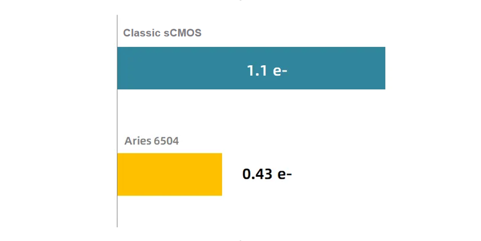
Deep-tissue scattering, rapid voltage dynamics, and the inherently low signal levels of some voltage indicators make weak-signal imaging particularly challenging. In many cases, weak signals lie at the noise floor, limiting both visibility and quantitative accuracy.
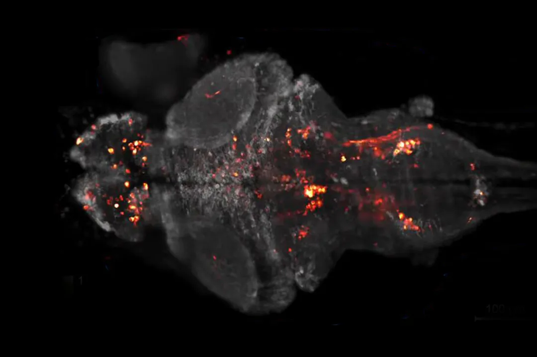
Figure 2: Calcium imaging for reference only
The Aries 6504 reduces read noise to 0.43 e⁻, approximately a 60% reduction from the previous model, achieving engineering-level sensitivity approaching the single-photon regime. This extends the lower bound of detectable signals and enhances stability and quantitative reliability, enabling a transition from “occasionally visible” to “consistently quantifiable” deep and weak-signal imaging. Under these conditions, imaging becomes primarily limited by the biological signal rather than the detector noise.
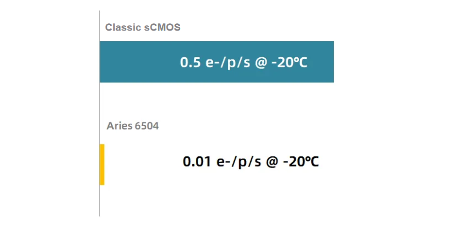
0.01 e⁻/pixel/s Dark Current — 50× Reduction
Improved Feasibility for Long-Exposure and Long-Duration Imaging
In in-vivo neuroscience, dark current is a key factor affecting long-exposure quality and long-duration recording stability. Over extended experiments, elevated dark current contributes to baseline drift and reduced quantitative consistency.
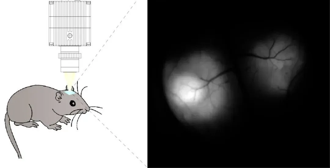
Figure 3: in-vivo neuroscience imaging for reference only
With dark current reduced to 0.01 e⁻/pixel/s at –20 °C, the Aries 6504 delivers a 50× improvement over the prior generation. This substantially enhances long-exposure performance and ensures image consistency during extended recordings. Reduced dark current also enables lower excitation light intensities, minimizing phototoxicity and photobleaching—critical for sensitive biological models and delicate experimental conditions.
Conclusion
Over the past decade, sCMOS technology has not only changed the scale at which research questions can be addressed, but also reshaped experimental design and deepened our understanding of how the brain operates.
We expect the Aries 6504, as a next-generation back-illuminated sCMOS camera, to continue advancing this trajectory—working in concert with emerging approaches such as adaptive optics, novel fluorescence probes, and computational imaging techniques (including deep-learning–based reconstruction). Together, these developments can help bring neuroscience closer to its long-standing aspiration: real-time, whole-system, cell-level observation of the living brain.
If you would like additional information on the Aries 6504 or to discuss its suitability for your applications, please feel free to contact us.
For a more detailed technical analysis of the Aries 6504 camera, refer to the product pre-release bulletin titled “Tucsen announces next-generation sCMOS camera improving speeds to 300fps and reducing read noise to a low of 0.43 electrons.”
Tucsen Photonics Co., Ltd. All rights reserved. When citing, please acknowledge the source: www.tucsen.com


