
 2022/08/24
Abstract
Electrical stimulation via invasive microelectrodes is commonly used to treat a wide range of neurological and psychiatric conditions. Despite its remarkable success, the stimulation performance is not sustainable since the electrodes become encapsulated by gliosis due to foreign body reactions. Magnetic stimulation overcomes these limitations by eliminating the need for a metal-electrode contact. Here, we demonstrate a novel microfabricated solenoid inductor (80 µm × 40 µm) with a magnetic core that can activate neuronal tissue.
Implantable micromagnetic stimulation (µMS)has several advantages over electrode-based stimulation. Improvements in nanofabrication technology have allowed us to create ultrasmall solenoids with magnetic cores that can generate larger magnetic fields while being completely encapsulated in a biocompatible coating. Novel microfabricated solenoid successfully activated neural tissue and therefore shows potential as a viable alternative to current neural interfacing tools for basic neuroscience and clinical applications, although further investigation is required.

Fig . a The setup used for measuring the magnetic flux density emitted by the novel microsolenoid using a custom-built system based on an NV diamond sensor. b The scan window is shown used in setup (a). c The setup used proof-of-concept of µMS using the micro/macrosolenoids in acute brain slices
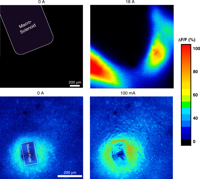
Fig . Epifluorescence micrograph of a brain slice from Thy1-GCaMP6s transgenic mice showing the change in fluorescence in response to µMS when using a (top) macrosolenoid and a (bottom) microsolenoid
Analysis of imaging technology
The Dhyana 400BSI camera was used to observe radio-fluorescence microscopic images of the brain slices of transgenic mice. It has good contrast and sensitivity, provides excellent quantum efficiency and low noise at UV wavelengths, and the high dynamic range 16-bit mode allows imaging of bright field and fluorescence even when the fluorescence signal is very low. The radiation changes of different sizes of solenoids to mouse brain slices can be seen intuitively in the image, so as to preliminarily determine the feasibility of the scheme. Submillimeter and millimeter coils convert the applied current into magnetic flux, which then induces an electric field gradient strong enough to move ions and push them to sense (or suppress) the neuron's response.
Reference source:
1.Khalifa, A., Zaeimbashi, M., Zhou, T.X. et al. The development of microfabricated solenoids with magnetic cores for micromagnetic neural stimulation. Microsyst Nanoeng 7, 91 (2021). https://doi.org/10.1038/s41378-021-00320-8


