

 2026/10/14
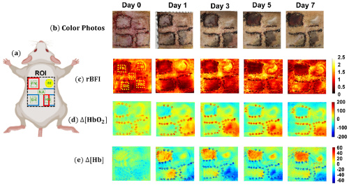
Researchers at the University of Kentucky developed a multi-wavelength speckle contrast diffuse correlation tomography (MW-scDCT) system to monitor blood flow and oxygenation in skin flaps after mastectomy-related reconstruction. In a rat study, combining these measurements distinguished necrotic from viable flaps with 80–95% accuracy. A Tucsen Dhyana 400BSI sCMOS camera captured the weak two-dimensional speckle signals used to generate the system’s blood-flow and oxygenation maps.
Why is skin-flap necrosis a concern after mastectomy?
Many women who undergo mastectomy later choose implant-based breast reconstruction. If the preserved skin flap does not receive enough blood and oxygen, part of the tissue may die. This complication is known as mastectomy skin flap necrosis (MSFN).
MSFN can impair wound healing, delay further cancer treatment, and compromise reconstruction outcomes. Earlier identification of poorly perfused tissue could help clinicians intervene sooner and better protect the reconstructed breast.
How does MW-scDCT assess flap health?
MW-scDCT uses near-infrared light to measure two key indicators of tissue health: blood flow and oxygenation. As light travels through tissue, moving red blood cells create rapidly changing speckle patterns. Analyzing these patterns provides information about perfusion.
By using multiple wavelengths, the system also estimates changes in oxygenated and deoxygenated hemoglobin. This allows researchers to map blood flow and oxygenation together, rather than relying on visual signs or a single measurement.
What did the study show?
The University of Kentucky team monitored rat skin flaps for seven days after surgery. Necrotic flaps showed lower blood flow and oxygenation, while viable flaps remained more stable or recovered over time.
When the researchers combined the relative blood-flow index with changes in oxygenated and deoxygenated hemoglobin, they could distinguish necrotic from viable flaps with 80–95% accuracy. Using any single parameter alone achieved only 40–70% accuracy.
Because these results come from an animal study, further validation is needed before clinical use. However, they support MW-scDCT as a promising approach for earlier, noncontact assessment of skin-flap viability.
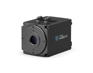
Why use the Dhyana 400BSI?
The MW-scDCT system depends on detecting faint, fast-changing speckle patterns formed by light scattered inside tissue. The Dhyana 400BSI served as the two-dimensional detector, recording the spatial speckle data required for blood-flow and oxygenation mapping.
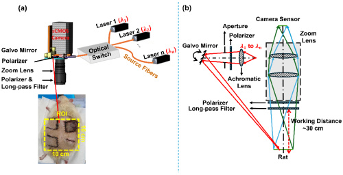
Its back-illuminated sCMOS sensor supports weak-light detection, while its 2048 × 2048 sensor captures detailed spatial information across the field of view. The study used a 5 ms exposure and a 25 Hz acquisition rate. The camera’s high-speed readout—up to 100 fps at 4.2 MP via Camera Link—provides additional capability for dynamic speckle imaging.
The Dhyana 400BSI V3 uses the GSENSE2020BSI sensor which gives high sensitivity sCMOS capabilities.
Can this approach support breast reconstruction?
If validated clinically, MW-scDCT could provide surgeons with noninvasive, objective information about skin-flap perfusion after mastectomy. Earlier detection of ischemic or hypoxic tissue may support timely intervention and help reduce reconstruction complications.
For this type of optical imaging, the Dhyana 400BSI provides the sensitivity, resolution, and speed needed to turn weak speckle signals into meaningful maps of tissue health.
FAQ
What is MW-scDCT?
MW-scDCT stands for multi-wavelength speckle contrast diffuse correlation tomography. It is a noncontact, dye-free optical imaging method that measures tissue blood flow and oxygenation by analyzing speckle patterns created by near-infrared light scattered through tissue.
Which camera was used in the MW-scDCT skin-flap study?
The published study used a Tucsen Dhyana 400BSI camera to capture two-dimensional speckle patterns for blood-flow and oxygenation analysis. Confirm the exact hardware version with the research team before making a model-specific claim.
How does the Dhyana 400BSI support speckle imaging?
Its back-illuminated sCMOS sensor detects weak scattered-light signals, while its 4.2 MP resolution and high-speed readout record the spatial and temporal speckle information required for tissue blood-flow and oxygenation mapping.
Explore More Application Stories
Discover how Tucsen scientific cameras support weak-light imaging, dynamic optical measurements, and advanced biomedical research applications.
Explore More Application Stories
Have a similar imaging requirement or need help selecting a camera for your system?
References
1. Robertson S, Jeevaratnam JA, Agrawal A, Cutress RI. Mastectomy skin flap necrosis: challenges and solutions. Breast Cancer: Targets and Therapy. 2017;9:141–148. https://www.dovepress.com/mastectomy-skin-flap-necrosis-challenges-and-solutions-peer-reviewed-fulltext-article-BCTT
2. Rab S, et al. Noncontact diffuse optical imaging of blood flow and oxygenation distributions in reconstructive rat skin flaps. Biomedical Optics Express. 2025;16(9):3740–3758.
https://opg.optica.org/boe/fulltext.cfm?uri=boe-16-9-3740
3. https://abs.amegroups.org/article/view/8262/html


